It is often said that prevention is better than cure. When it comes to eye health this is no different, as around 90% of vision impairment in Australia is preventable if detected early. Queensland Eye and Retina Specialists are strong advocates of preventative eye care, and provide their team of highly skilled ophthalmologists with high quality equipment to detect any early signs of disease.
One such device is the Nikon Optos, which is the only machine currently available to take an anatomically correct image of up to 82% of the retina in a single image. This is particularly important for discovering and diagnosing pathology that only presents itself in the peripheral retina. The Nikon Optos also allows for a number of imaging modalities, including red-green, red-free, autofluorescence and both fluorescein and indocyanine green angiography. A more in depth discussion of fluorescein angiography and its role in the management of numerous eye conditions can be found in our blog ‘Fluorescein Angiography and the Nikon Optos’.
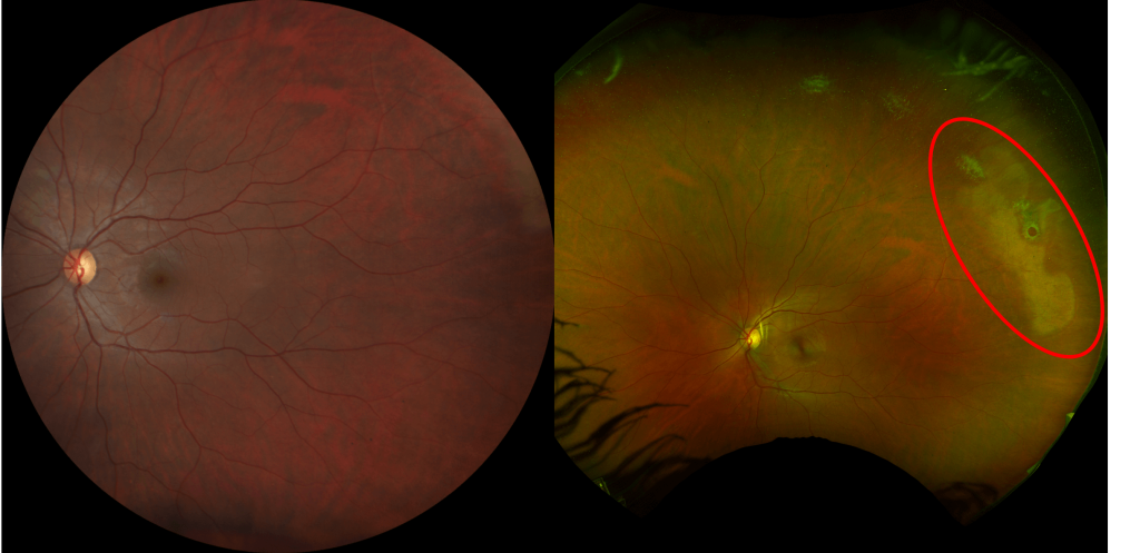
The image above on the left demonstrates a 45° view of the retina, taken using a high-definition fundus camera. The retina of the same patient can be seen on the right, however, this time the image was taken using the Nikon Optos. This reveals previously unseen retinal changes, in this case lattice degeneration and atrophic retinal holes, which can be missed using traditional imaging techniques.
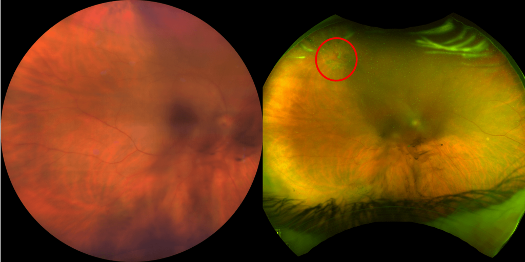
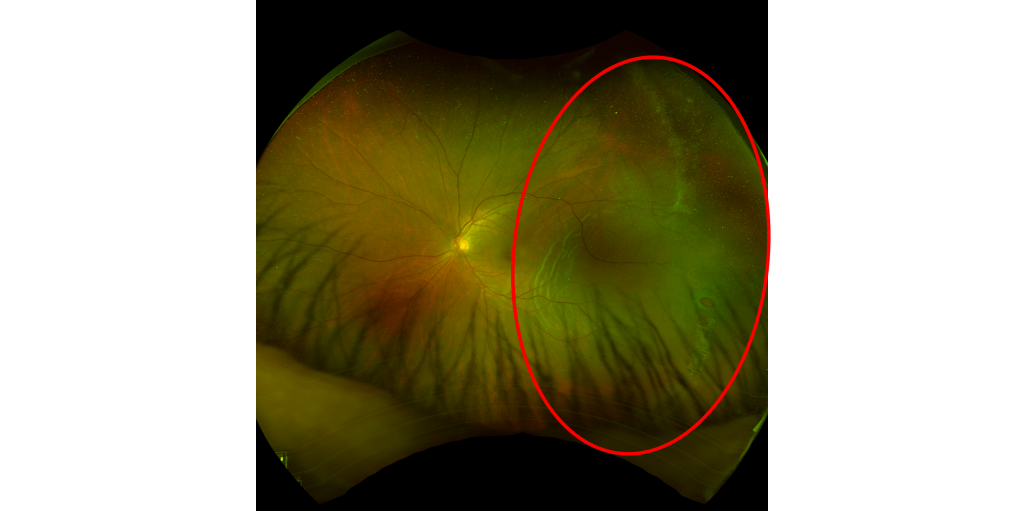
Retinal tears and detachments are among other sight-threatening eye conditions where the Nikon Optos provides invaluable insight. One such example is the patient above who presented with sudden onset floaters and flashes and blurry central vision. The conventional fundus image on the left is unable to determine the cause, whilst the Optos image on the left shows a horseshoe retinal tear with avulsed vessels causing vitreous hemorrhage. If diagnosed and treated in a timely manner, it is possible to avoid a retinal tear turning into a retinal detachment, which can cause permanent vision impairment. The image below illustrates a retinal hole which progressed into a retinal detachment.
Referring patients to Queensland Eye and Retina Specialists means that they will get a thorough examination with multiple different diagnostic machines. Use of multi-modal imaging increases our ophthalmologist’s ability to detect, diagnose and treat conditions, even in the peripheral retina. Our dedication to utilising cutting-edge, advanced equipment underscores our pursuit of excellence in the diagnosis and treatment of our patients.