The multi-modal imaging capabilities of the Nikon Optos allow the ophthalmologists at Queensland Eye and Retina Specialists to diagnose and treat a number of potentially sight-threatening eye conditions. The fundus fluorescence angiography (FFA) and indocyanine green angiography (ICGA) modalities on the Nikon Optos play a significant role in assessing the blood supply to the retina and choroid. These tests are particularly important in conditions which affect retinal vasculature, such as; macular oedema, diabetic retinopathy, vein occlusion, Eale’s disease etc.
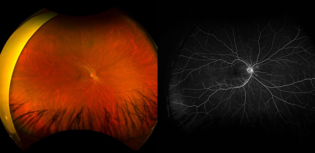
In these procedures a dye, which fluoresces under exposure to certain wavelengths of light, is injected intravenously, usually into a vein in the arm or hand. The ophthalmologist then uses a camera with specialised filters to capture the light emitted from the fluorescein or indocyanine molecules which are flowing through the blood vessels in the back of the eye. A comparison between a standard Optos image (left), and a fluorescence angiogram (right) on the same patient can be seen below, illustrating the enhanced visualisation of retinal vasculature available with fluorescein angiography.
One condition, in which FFA provides the ophthalmologists of QERS with vital insights, is diabetic eye disease. Currently FFA is the gold standard imaging technique to assess the retinal vasculature for signs of non-proliferative and/or proliferative diabetic retinopathy. In cases of diabetic retinopathy there can be a number of signs which can be difficult to appreciate using standard fundus photography. A number of examples below will demonstrate these signs.
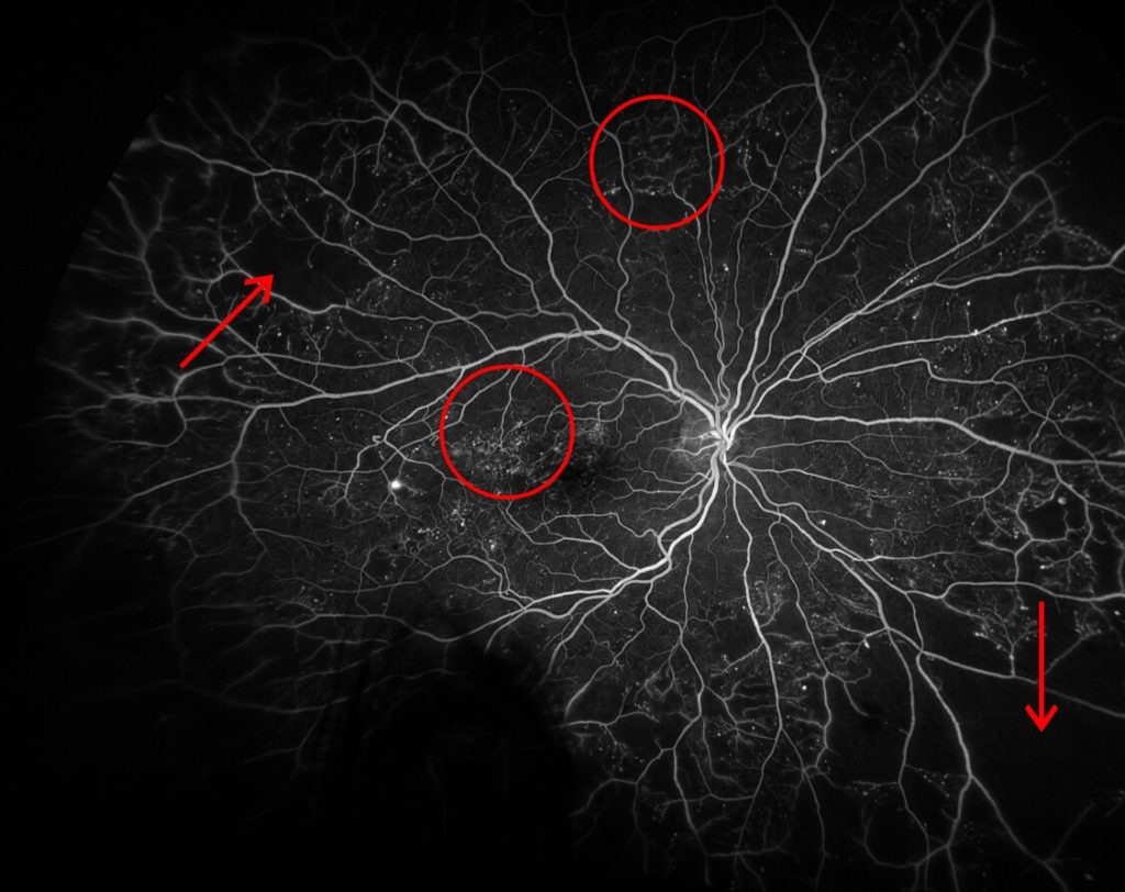
The above image demonstrates changes associated with early proliferative diabetic retinopathy. Numerous examples of hypofluorescent non-perfused/ ischemic areas can be seen in the peripheral retina (indicated by arrows). Scattered hyperfluorescent micro-cystic changes and numerous intraretinal retinal microvascular anomalies (IRMA) can be seen in the circled areas.
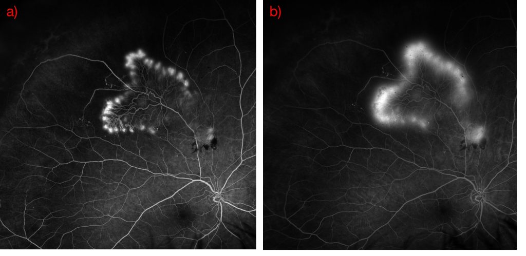
A further unique capability of FFA is its ability to identify vascular leakage, a sign which is of particular importance in several retinal diseases. One example can be seen below, where a frond of diabetic neovascularization elsewhere (NVE) is seen in the superior retina. The FFA images below demonstrate early hyperfluorescence of the frond of new vessels (a) and subsequent late leakage of the vessels (b). Untreated, these NVE may lead to further complications, including diabetic macula edema, further neovascularization or tractional retinal detachment. Evidence of vascular leakage such as this may prompt an ophthalmologist to recommend a treatment of sectoral panretinal photocoagulation (PRP). This treatment causes destruction of the peripheral retina, with the aim to reduce triggers for new vessel formation and potentially regress current NVE.
Access to this modality of imaging is vital in the diagnosis and management of diabetic eye disease, along with a number of other retinal diseases. At Queensland Eye and Retina Specialists we specialise in the diagnosis and treatment of diabetic retinopathy. All the Ophthalmologists are trained in medical retinal treatment (i.e. Anti-VEGF Intravitreal injections) and Dr Sean Cheng and Dr Rajan Patheja are further specialised in surgical treatment (i.e. Vitrectomy, Panretinal Photocoagulation).
Queensland Eye & Retina Specialists
accepts referrals via email, Oculo, Medical Objects and fax.