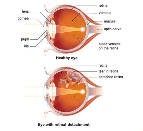
What is Retinal Detachment?
The retina is a light sensitive layer which lines the inner surface of the back of the eye and acts like a camera film to capture light. The light that reaches the retina forms an image which is sent to, and interpreted by, our brain – this is how we see.
When the retina is separated from its normal position – similar to wallpaper peeling off a wall – it is known as a retinal detachment. It is potentially a sight-threatening emergency and must be reviewed by an ophthalmologist within 24-48 hours, as it may lead to permanent vision loss if not properly managed in a timely manner.

Symptoms and Risks
Symptoms
Symptoms of retinal detachment may include:
- Sudden onset of seeing floaters (dark spots, ‘blobs’, wiggly lines) and/or light flashes
- Some vision impairment described as a dark curtain or shadow
- Depending on where the detachment is, the patient may experience sudden central vision loss
Causes and Risks
There are many causes and risk factors, including:
- A tear or a hole in the retina
- Extreme near sightedness (Myopia)
- Eye trauma
- A family history
- A previous diagnosis
- Complication from previous eye surgery
Book a QERS Consultation
Treatment
Options
Retinal detachment can only be treated by surgery. If treated early, there is a higher chance of regaining lost vision. Common procedures include scleral buckle, vitrectomy, or a combination of both.
Scleral buckle involves placing a silicone band around the circumference of the eyeball. This pushes the wall of the eyeball (choroid) into contact with the retina and can stay permanently in place.
Vitrectomy involves removing the vitreous jelly within the eyeball and replacing it with air, gas, or silicone oil to support the retina flat against the back of the eye, allowing the detached retina to heal. The amount of vision recovered will depend on the extent and duration of the retinal detachment, whether the central vision (macula) was involved, and any other pre-existing eye conditions. You may require subsequent surgeries and this will be discussed with you.
Immediately after surgery, specific posturing of the head may be required to assist the gas or air bubble in pushing the retina back into place. Detailed instructions will be provided if you are required to posture for recovery.
Your Journey
At Queensland Eye & Retina Specialists, our clinician will use state-of-the-art precision equipment to examine your eye. Our specialist surgeon will then discuss an individualised treatment plan for your best visual outcome.
As with all surgery, there are some risks involved which will be discussed with you at your consultation.
More Information
For more information on retinal detachment, head to one of the links below

