12 Cases of Christmas
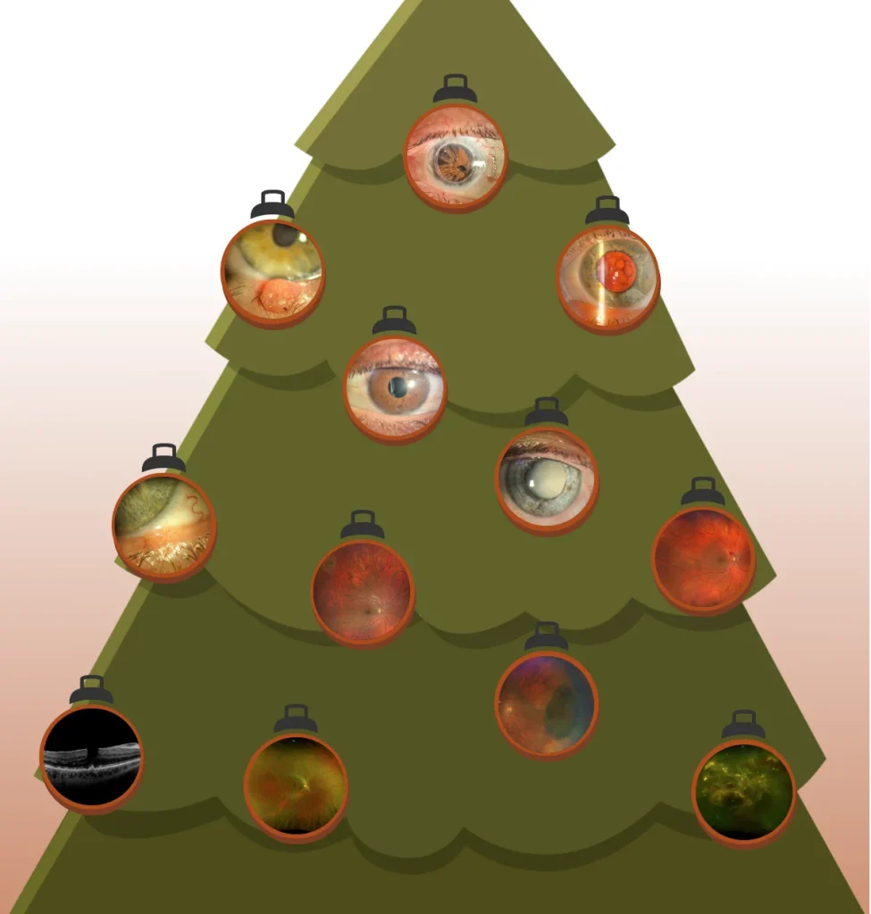
Do you know what the conditions of these 14 eye-baubles are?
There are 14 eye-baubles telling the stories of 12 different case studies.
When you look at the pictures, are you able to describe the condition and what the treatment may have been? Take a look and then scroll over eye-bauble to discover the case study details.
Bilateral corectopia and dyscoria associated with Axenfeld-Rieger syndrome. This autosomal dominant genetically inherited condition often results in anterior segment developmental abnormalities. 50% of patients diagnosed with Axenfeld-Rieger syndrome will develop glaucoma. This patient also had a penetrating keratoplasty (PK) or corneal transplant in the left eye.
Bilateral corectopia and dyscoria associated with Axenfeld-Rieger syndrome. This autosomal dominant genetically inherited condition often results in anterior segment developmental abnormalities. 50% of patients diagnosed with Axenfeld-Rieger syndrome will develop glaucoma. This patient also had a penetrating keratoplasty (PK) or corneal transplant in the left eye.
A left lower lid lesion which had previously been diagnosed as a benign papilloma. On clinical examination there were no suspicious features, although the only way to definitively diagnose is via biopsy. The histopathology report following biopsy identified the lesion as an inverted follicular keratosis. This is a benign tumour, more commonly occurring on the heads of older people.
Bilateral posterior-subcapsular cataracts causing reduced best corrected visual acuity of RE: 6/9 and LE 6/21. Following cataract surgeries, with EDOF IOLs, unaided visual acuity improved to 6/4.5 in both eyes with near acuity of N8 unaided at 40cm.
A subluxated 3-piece IOL with one haptic extending into the anterior chamber. Following vitrectomy, removal of the subluxated IOL and implantation of a scleral fixated IOL, the gentleman’s visual acuity improved to 6/7.5.
A dense, mature white cataract in the right eye. The cataract was thought to be related to high dose steroid use for severe Graves’ ophthalmopathy (aka thyroid eye disease). Surgery on a cataract of this density is associated with increased rates of intraoperative complications, such as posterior capsule tear, zonular dehiscence or dropped lens material. Following successful surgery, visual acuity improved to 6/6 unaided.
The patient had noticed that her left eye felt a bit strange recently. Upon visiting her optometrist, a small lump was evident on the left lower lid. It had been increasing in size in the past 2 weeks. The patient had a history of a number of previous squamous cell carcinomas (SCC) and basal cell carcinomas (BCC). Although clinically the spot appeared to resemble a papilloma, a biopsy was taken.
The patient had attended their optometrist for a routine eye assessment. He was moderately myopic (~-5.00D) and had a history of longstanding floaters and intermittent flashes in the right eye. On examination through dilated pupils a small operculated hole was identified, and the decision was made to treat with barrier retinal laser to reduce the likelihood of the operculum progressing to a retinal detachment.
The patient had noticed black blurry lines in her central vision, which prompted her to see her optometrist. Upon dilation, a retinal hemorrhage and cotton wool spot were seen superotemporal to the optic nerve head. Cotton wool spots are believed to occur following ischemic changes, usually from retinal arterial occlusion. In a high number of cases, a single cotton wool spot may be the first sign of hypertensive retinopathy or diabetes. Management for this patient included Alphagan P eye drops twice daily for its potential ischemic neuroprotective benefits, and a cardiovascular risk screening, looking for underlying causes such as hypertension, diabetes and hypercholesterolaemia.
A large CHRPE (congenital hypertrophy of the retinal pigment epithelium) in the nasal fundus of a patient. The CHRPE had been picked up during a routine optometric assessment and the patient was asymptomatic. CHRPEs are a localised malformation of RPE cells, which are usually present at birth. They are typically benign and require no treatment, however the atypical variant has an association with a familial adenomatous polyposis (FAP).
The patient had noticed a general very gradual reduction in visual quality over the past two years, which was particularly noticeable when reading. His visual acuity was still well maintained with a mild myopic prescription allowing him to achieve 6/6 vision in both eyes. Upon investigation with OCT imaging, there was mild dry age-related macular degenerative changes with drusen and epiretinal membrane with a pseudo-hole appearance. These changes were present in both eyes. Due to the well-preserved visual acuity the patient opted for conservative management and reassessment in 6 months’ time.
The patient had noticed for about one-week spots in her vision in the right eye without any pain. She had longstanding “floaters” which moved around as her eye moved, but she felt the new spots were different. Instead of moving around, they were fixed in her visual field and covered letters when she was trying to read with her right eye alone. Based on the appearance of the fluorescein angiogram she was diagnosed with multiple evanescent white dot syndrome (MEWDS). She fit the demographic of patients most likely to be diagnosed with MEWDS, a myopic female between 15 and 50 with an associated viral prodrome. Fortunately, MEWDS is self-limiting and nearly all patients will recover normal vision in 3-9 weeks.
The patient had noticed for about one-week spots in her vision in the right eye without any pain. She had longstanding “floaters” which moved around as her eye moved, but she felt the new spots were different. Instead of moving around, they were fixed in her visual field and covered letters when she was trying to read with her right eye alone. Based on the appearance of the fluorescein angiogram she was diagnosed with multiple evanescent white dot syndrome (MEWDS). She fit the demographic of patients most likely to be diagnosed with MEWDS, a myopic female between 15 and 50 with an associated viral prodrome. Fortunately, MEWDS is self-limiting and nearly all patients will recover normal vision in 3-9 weeks.
This patient from Papua New Guinea attended Queensland Eye and Retina Specialists after seeing a local ophthalmologist. He had previously noticed an acute painless loss of vision in the right eye three years earlier. His visual acuity in the RE was no perception of light (NPL) and a marked right RAPD was observed. He was diagnosed with end-stage Coats’ disease, a telangiectatic neovascular disease of the retina, which most commonly affects one eye of young males. At this stage there is no prognosis for visual improvement and no recommended treatment unless complications arise, such as neovascular glaucoma or phthisis bulbi. It is important to distinguish between Coats’ disease and retinoblastoma.